![]()
A smarter way to “see” inside the body and enhance cancer diagnosis
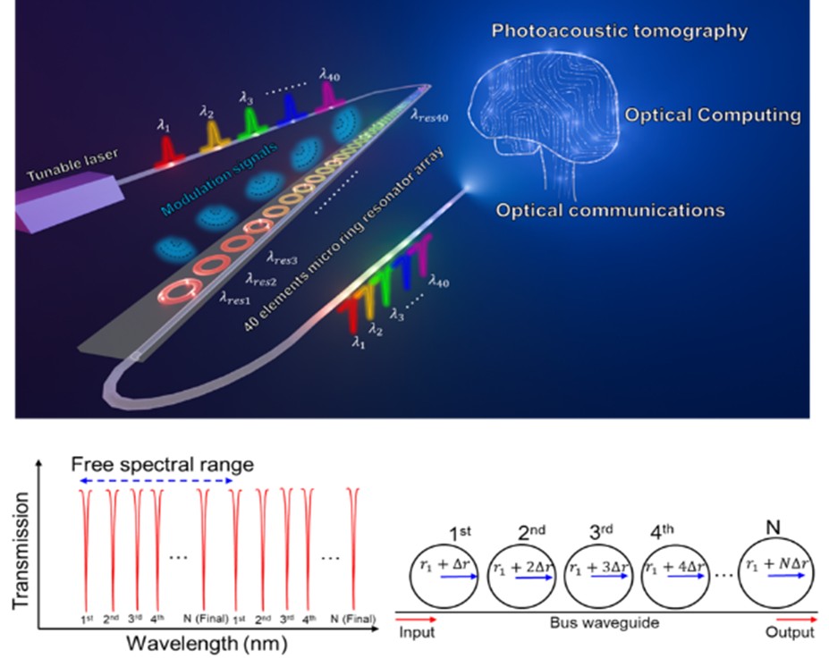
CHINA, June 22, 2026 /EINPresswire.com/ — A team of researchers has developed a new type of imaging sensor that uses both light and sound to create detailed pictures of biological tissues. This marks the first successful use of an array of over 40 microscopic, polymer-based “microrings” to capture these types of images. It can track oxygen levels, blood flow, and how dense blood vessels are. Therefore, it is much more effective at spotting the early signs of cancer.
A team of engineers from the University of Michigan led by Professors Xueding Wang, Guan Xu, and L. Jay Guo collaborated to advance optical detection of ultrasound and demonstrate photoacoustic tomography (PAT) using optical arrays. Recent advances in biomedical imaging have significantly improved the ability to visualize biological structures inside the body. However, many imaging techniques face trade-offs between resolution, sensitivity, depth, and functional contrast. PAT has emerged as a promising hybrid imaging modality that combines the strengths of optical contrast and ultrasound imaging. By using short laser pulses to generate ultrasound waves through light absorption in tissue, PAT can reveal optical absorption contrast at depths beyond those accessible by purely optical techniques. This capability makes it particularly attractive for applications such as cancer detection, where changes in blood vessel density, hemoglobin concentration, and tissue architecture are important diagnostic indicators. The article was made available online on March 30, 2026, and was published in Volume 9, Issue 6, of the journal Opto-Electronic Advances, on June 07, 2026.
Despite its promise, the broader clinical adoption of PAT depends heavily on the performance and scalability of ultrasound detection technology. Conventional ultrasound transducers are typically based on piezoelectric materials, which can limit bandwidth, miniaturization, and integration density. Optical ultrasound sensors, particularly microring resonators, have emerged as an alternative approach offering high sensitivity, wide bandwidth, and compatibility with photonic integration. However, scaling such optical sensors into large arrays with consistent performance has remained a technical challenge.
This work demonstrates a significant advance in this area by introducing a polymer-based microring resonator array containing more than 40 elements, fabricated using nanoimprint lithography. Nanoimprint lithography is a scalable and cost-effective nanofabrication method capable of producing nanometer-scale features over large areas. By precisely controlling the radius of each microring resonator at the nanometer scale, the team achieved multiple distinguishable resonances within a very narrow spectral window. This level of control enables the creation of dense optical sensor arrays while maintaining high optical quality factors, which are essential for sensitive ultrasound detection.
When integrated into a PAT system, the microring array demonstrated a broad acoustic detection bandwidth exceeding 170 MHz and achieved a fine spatial resolution on the order of tens of micrometers. Such broadband detection is particularly important because higher-frequency acoustic components carry information about smaller tissue structures. In practical demonstrations, the system was used to image ex vivo mouse prostate tissue. The resulting images showed strong correspondence with known biological structures, including blood vessel regions. Furthermore, spectral analysis of the photoacoustic signals allowed differentiation between normal and cancerous prostate tissues, suggesting the platform’s potential for functional and pathological tissue assessment.
Beyond the immediate imaging results, the broader significance of this work lies in its demonstration of a scalable fabrication strategy for high-performance optical ultrasound arrays. The use of polymer materials offers additional flexibility and potential for integration into compact photonic systems. Compared to traditional fabrication methods, nanoimprint lithography provides a pathway toward cost-effective mass production of sensor arrays, which is a key requirement for future clinical translation and widespread deployment.
In a broader technological context, microring resonator arrays fabricated by nanoimprinting may also find applications beyond biomedical imaging, including optical communication systems and integrated photonic circuits, where compact and high-performance resonators are essential components. By bridging scalable nanofabrication with advanced optical sensing and imaging technologies, this work contributes to the ongoing effort to develop next-generation diagnostic tools that combine high resolution, functional contrast, and practical manufacturability.
***
Reference
Title of original paper: Imprinted high-Q polymer micro-ring resonator array for high-resolution photoacoustic tomography
Journal: Opto-Electronic Advances
DOI: 10.29026/oea.2026.250215
About Prof. Xueding Wang’s team from the University of Michigan
The Optical Imaging Lab, directed by Professor Xueding Wang at the University of Michigan, focuses on advancing optical and ultrasound imaging technologies for biomedical applications. One area of particular interest is photoacoustic imaging, which combines light and sound to achieve highly sensitive, high-resolution imaging of deep biological samples. This technique holds promise for diagnosing conditions like inflammatory arthritis, breast cancer, prostate cancer, liver diseases, eye disorders, bowel diseases, bladder cancer, and brain disorders. Additionally, the lab explores nanoparticle agents made from materials, such as gold or organic hydrogels, which can enhance diagnostic imaging or deliver drugs.
About Prof. Guan Xu’s team from the University of Michigan
The Biomedical Imaging and Biomechanics Lab directed by Professor Guan Xu is a biomedical imaging and biomechanics research group at the University of Michigan and the Kellogg Eye Center. Their goal is to implement state-of-the-art optical, ultrasound and photoacoustic imaging, and biomechanical analysis technologies to the understanding and diagnosis of diseases.
About Prof. L. Jay Guo’s team from the University of Michigan
The Guo Research Lab focuses on nanomanufacturing and photonic/optoelectronic devices, with nanoimprint lithography as a versatile fabrication technology. The polymer microring ultrasound detector was first developed in the lab over two decades ago, and its performance has been improved ever since. Apart from opto-acoustic sensors and actuators, the lab also developed flexible transparent conductors, structural colors and AI-assisted design, hybrid photovoltaics and photodetectors, and nanomanufacturing technologies. Through these efforts, the lab continues to bridge advanced nanomanufacturing with modern photonic system applications.
Funding information
The study received financial support from the National Cancer Institute (grant numbers: R01CA278833 and 5R37CA22282903) and the National Institute of Diabetes and Digestive and Kidney Diseases (grant number: 1R01DK12568701).
Siyi Ma
Institute of Optics and Electronics
oej_publishing@ioe.ac.cn
Legal Disclaimer:
EIN Presswire provides this news content “as is” without warranty of any kind. We do not accept any responsibility or liability
for the accuracy, content, images, videos, licenses, completeness, legality, or reliability of the information contained in this
article. If you have any complaints or copyright issues related to this article, kindly contact the author above.
![]()
Media gallery